Life and Material Sciences
Broadband, High-Brightness Light in Materials Science & Life Science Applications
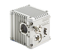
High Performance Analytical Spectroscopy is widely used in the materials and life sciences to analyze the molecular composition of complex materials. To achieve the most precise analysis, wavelengths that cover the range from the deepest ultraviolet through visible and into the near infrared. Traditionally, combinations of deuterium lamps and tungsten halogen lamps have been used. These lamps suffer from short operating life, low brightness and limited spectral range. Energetiq’s EQ-99X and EQ-77 Laser-Driven Light Sources offer much higher brightness across the whole spectrum, from 170nm to 2100nm (UV-Vis_NIR) and an order of magnitude longer life, enabling a new class of instruments capable of analyzing much smaller samples, with high precision and throughput.
Surface Science techniques in the materials sciences, such as Photoemission Electron Microscopy (PEEM), can take advantage of the high brightness and deep ultraviolet output of LDLS sources such as the EQ-77. In PEEM applications, the high photon energy, i.e. short wavelength, light must be focused to a small spot on the sample, perhaps 100µm in diameter. Usually, this must be accomplished at a significant working distance and through a vacuum window in the PEEM instrument. Traditionally used Xe and Hg arc sources have little output in the 200nm wavelength range, and their large plasma size, in the millimeter range, makes imaging the source on the sample an inefficient process. By contrast, the ~150um diameter high-brightness plasma in the EQ-1500 allows straightforward and efficient re-imaging of the source onto the sample, even with a long working distance. The high output of short wavelengths, usually selected from the broadband LDLS spectrum by narrow bandpass filters, has been demonstrated to produce PEEM images with significant intensity and contrast.
Biological Imaging - The ability to see the structure of living organisms and to analyze the relationship between form and function is key to biological studies. However, living organisms are dynamic structures, and biological processes that occur over time such as protein changes, cell division, embryo development, or neural activity necessitate the visualization of structures in multi-dimensions. With the development of advanced biological imaging technology it is now possible to observe molecular and cellular interactions that underlie essential functions within living cells and tissues as they take place. The Energetiq EQ-99X and EQ-77 Laser-Driven Light Source are ideal tools to use for these particular studies due to the high resolution, power, and brightness of the light generated. By utilizing DUV light as the light source, the nanostructure of living cells and tissues can be directly observed with extremely high resolution, without the extreme purchasing and operating costs associated with the use of conventional lasers.
Request More Information
Are you looking for more information on this product family? Fill out the form to request a quote, demo, or additional technical information.
Soft X-Ray Light in Life Sciences Applications
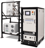
Soft X-Ray Microscopy and Soft X-Ray Microbeams - Soft x-ray light for microbeam studies and microscopy can readily be found in national lab synchrotron facilities. However, this limits the availability of soft x-rays to those researchers who are able to obtain beam time and who are able to bring their experiments to the synchrotrons. With its novel plasma approach, Energetiq’s compact EQ-10SXR Soft X-Ray Light Source enables soft x-ray microbeam and microscope experiments to be carried out in any typical biological research laboratory.
Microbeam studies of the effects of ionizing radiation on biological cells have long been performed using particle accelerators. The limitations of charged particle beams such as alpha particles include difficulty in focusing the beam to below one micrometer in diameter. Soft x-rays provide similar ionizing radiation effects on biological samples, but are readily focused to very small spot size using Fresnel zone-plate lenses. In principle, beams significantly less than 100nm in diameter can be produced using such optics. This allows the targeting of sub-cellular structures within cells with a controlled soft x-ray beam, to evaluate the response of such structures to ionizing radiation. Applications include the effects of environmental radiation, and the mechanisms of radiotherapy for cancer and the like.
In soft x-ray microscopy, images produced with soft (low energy) x-ray beams have 10 times the resolution of images obtained with the best visible light microscopes. In addition, studies can be done on living cells without destroying them. Soft x-ray microscopy can make a major contribution to the understanding of cell function and structure. The Energetiq EQ-10SXR light source has been shown to produce sufficient 2.88nm light to allow production of high resolution images in a practical time.


